Mastering the Art of Spore Staining: A Crucial Microbiology Skill
News 8 4 月, 2025
Spore staining is a vital microbiological technique for identifying spore-forming bacteria that are otherwise difficult to visualize using standard Gram staining. While Gram staining helps differentiate bacteria as Gram-positive or Gram-negative, it often fails to stain bacterial spores effectively. Under a microscope, these spores appear as hollow, unstained structures, either free or at one end of the bacterium.
What Are Bacterial Spores?
Bacterial spores are dormant, highly resistant structures formed by genera such as Bacillus and Clostridium. They are spherical or oval in shape, possess thick walls, and have low water content. These properties make them highly resilient to heat, radiation, and chemical disinfectants. Though spores do not reproduce (each vegetative cell forms only one spore), their exceptional resistance poses a significant challenge in food sterilization, often leading to spoilage.
Why Spore Staining Matters
To visualize these tough microbial structures, specific staining methods are required. One of the most effective techniques is the Schaeffer-Fulton method, which uses malachite green to stain spores and safranin as a counterstain for vegetative cells.
Schaeffer-Fulton Spore Staining Protocol
Step-by-Step Guide:
- Smear Preparation: Choose spore-forming bacteria in the proper growth phase. Prepare a smear on a glass slide and fix it by heat.
- Primary Staining: Apply 5% malachite green to the smear. Heat over steam for 5 minutes, allowing vapor formation but avoiding boiling. Replenish the dye as needed.
- Cooling and Washing: Allow the slide to cool, then rinse with water until the runoff is colorless.
- Counterstaining: Apply 0.5% safranin for 1 minute to stain the vegetative cells.
- Final Wash and Observation: Rinse, dry, and observe under a microscope.
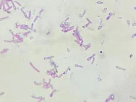
Expected Results:
- Spores appear green
- Vegetative cells appear red
Why This Method Works
Malachite green weakly binds to bacterial cell walls and spore coats. Washing with water easily removes the dye from vegetative cells but not from the spore core, which retains the green color. Safranin then stains the vegetative cells red, allowing clear differentiation under the microscope.
Key Tips for Successful Spore Staining
- Use the Right Growth Stage: Select bacteria in the appropriate phase where spores and vegetative cells coexist.
- Avoid Overheating: Excess heat during fixation (>200°C) can deform cells.
- Sufficient Heating of Malachite Green: Maintain heat without boiling to drive the dye into the spores.
- Prevent Drying During Heating: Keep the smear moist by adding dye as needed to avoid background staining and unclear visuals.
Conclusion
Mastering the spore staining technique is essential for microbiologists working with resilient spore-forming bacteria. With the right approach and attention to detail, this method enables clear visualization of spores, contributing to better bacterial identification and contamination control.